McGill Materials Services | McGill University
Material Testing and Characterization Service
The resources available to get what you need done, done

Discover More

McGill Materials Services


Dr. Florence Paray is the Director and Coordinator of the McGill Materials Services on behalf of the Department of Mining and Materials Engineering, McGill University.

Please contact us for service costs, an appointment, a special project or any other need you may have.

Dr. Florence Paray
Tel.: (514) 398-3590
Fax: (514) 398-4492

Analytical Labs


Aleksandra Djuric - Technician (514) 398-2610
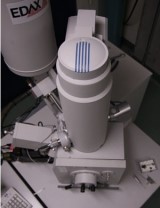
Equipment:


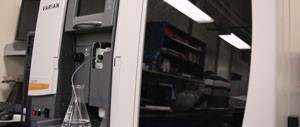
Atomic Absorption Spectroscopy

- Perkin Elmer Atomic Absorption Spectrometer 311. Highly sensitive instrument (ppm level).

- Varian AA 240 FS Atomic Absorption Spectrometer Fully automated PC-controlled double-beam atomic absorption spectrometer with the fast sequential operation for multi-element (4) flame AA determination.


UV Spectroscopy

- Perkin Elmer Lamda 20 UV/Vis Spectrometer

Ion Chromatography

- Dionex DX-00 Ion Chromotography

Laser Scattering Particle Size Distribution Analysis

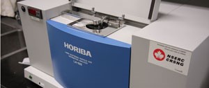
- Horiba Laser Scattering Particle Size Analyzer

- Size range from 0.020 to 2000 microns.


Particle Analysis

- Micrometric TriStar Surface area and porosity analyzer

- BET analysis of up to 3 samples at the time

Electron Microscopy Labs


Dr. Florence Paray - 514 398-3590

Equipment:

The Department of Mining and Materials Engineering has three exceptional research tools with its Scanning Electron Microscopes (SEM).

Hitachi SU-3500 Variable Pressure - SEM

- Resolution SE: 3nm at 30kV (high vacuum mode), 7nm at 3kV (high vacuum mode)

- Ultra variable pressure, BSE, SDD EDS and EBSD detectors

- The BSE detector allows obtaining a quantitative three-dimensional maps of the surface of a specimen, allowing to quantify complicated and/or rough surfaces

- Stereoscopic image function


Hitachi Cold FE SU-8000 SEM

- 0.5 nm resolution at 30 kV and 2 nm resolution at 0.2 kV.

- 7 Detectors including a secondary electron (SE) lower detector, a SE/BSE upper detector and SE/BSE top detector with energy filtration, a five quadrant BSE detector, a STEM detector for bright/dark field imaging a SDD EDS Detector and an EBSD camera.


Hitachi Cold FE SU-823000 SEM

- 0.5 nm resolution at 30 kV and 3 nm resolution at 0.05 kV.

- Secondary electron (SE) lower detector, SE/BSE upper detector, SE/BSE top detector with energy filtration of BSE electrons, five quadrant BSE detector, STEM detector for bright/dark field, and SDD EDS detector.

Facility for Electron Microscopy Research (FEMR)


Facilities for Electron Microscopy Sample Preparation, Scanning Electron Microscopy (SEM), and Transmission Electron Microscopy (TEM) for both biological and material sciences applications.

The Department of Mining and Materials Engineering is hosting some sample preparation equipment and the following Scanning Electron Microscopes:

Equipment:


FEI Quanta 450 Environmental Scanning Electron Microscope (FE-ESEM)

EDAX Octane Super 60 mm2 SDD and TEAM EDS Analysis System

FEI Inspect F-50 FE-SEM

EDAX Octane Super 60 mm2 SDD and TEAM EDS Analysis System and iXRF XBeam XRF

Materials and Characterization Laboratories


Dr. Florenca Paray (514) 398-3590

- Sample preparation

- Light Optical Microscopy

- Stereomicroscopy

- Quantitative metallography

- Image analysis

Robert Paquette, Technician (514) 398-5587

- Heat treatment

- Hardness testing (Vickers, Knoop, Rockwell, Brinell, Microhardness).

- Mechanical Testing(Charpy, tensile, three points, four points bending, etc.)


X-Ray Diffractometers


Philips PW 1710 Powder X-Ray Diffractometer

- Identification of constituents and crystalline structure of minerals, corrosion products, and inorganic materials.

- Qualitative and semi-quantitative analysis of crystalline phases in a sample

Monique Riendeau, Technician (514) 398-2610

Bruker D8 Discovery X-Ray Diffractometer

- High-tech, non-destructive technique for analyzing a wide range of materials, including fluids, metals, minerals, polymers, catalysts, plastics, pharmaceuticals, thin-film coatings, ceramics, bio-materials, and semiconductors.

- Use of radiation from a Co-tube for texture and residual stress investigations.

- Typical XRD diffraction analyses.

Dr. Florence Paray (514) 398-3590

McGill Institute for Advanced Materials (MIAM)


MIAM was established by the Faculties of Science and Engineering to act as a focal point for research into all forms of advanced materials. MIAM brings together diverse research facilities housed in various departments and faculties at McGill.

Subunit in the Department of Mining and Materials Engineering:

- Static Imaging MicroCT (MCT): Skyscan 1172

- X-Ray Diffractometry (XRD): D8 Discover Bruker (Cu source)

- X-Ray Photoelectron Spectroscopy (XPS): Thermo Scientific K-Alpha

Dr. Lihong Shang (514) 398-2541

MIAM Website